|
Working Wonders
Outside the Treatment Box with
CranioSacral Therapy |
 CranioSacral
Therapy (CST) is often misunderstood in the scientific world of
evidence-based medicine. The problem is partly because of different
interpretations of what CST entails and partly because of a lack of
understanding of various researchers as to the core principles involved in
the evaluation and treatment of the craniosacral system (CSS).12345
Many healthcare practitioners study CST and include it in their skill set6,
and patients often seek out CST when traditional medical practices fail.7
Although not a panacea, CST experiences have been catalogued in a book
titled Working Wonders.8 Wonder is defined as a cause of
astonishment at something new to one’s experience.9 CST was
certainly new to my experience over 35 years ago and to this day. I continue
to marvel in astonishment at the results that can be achieved through this
light-touch therapy. Although wonder is not commonly associated with
evidence-based medicine, it does indeed fuel scientific inquiry. Ralph Waldo
Emerson referred to wonder as “the seed of our science,”10 while
Abraham Heschel described wonder as the “the root of knowledge.”11
Experiencing wonder invites the question “why?”—which leads to scientific
inquiry.
Anatomical root of CranioSacral Therapy
To explore the wonder of CST, a brief overview of the anatomy of the CSS is
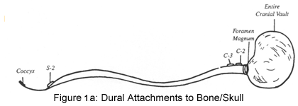
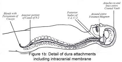
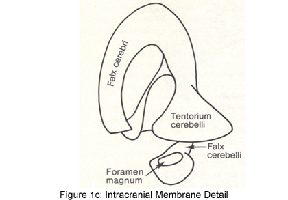
in order. The cranium is lined with dura mater, which not only encircles the
inner surfaces of the cranial bones but also folds in on itself. This
creates the falx cerebri, tentorium cerebelli and the falx cerebelli,
otherwise known as the intracranial membrane (ICM). The firm attachment of
the falx cerebelli at the foramen magnum of the occiput continues inferiorly
with attachments on the posterior bodies of C1 and C2. It continues in the
inferior direction without any attachments until it anchors at the S2
segment as the pia portion of the filum terminale within the sacral canal.
It exits out of the sacral canal and continues as the external dural segment
of the filum terminale blending with the periosteum of the coccyx (figures
1a,b,c).12,13 In addition, the dura mater extends out through the
intervertebral foramina with the spinal nerves as the dural sleeves. The
dural sleeves attach on the vertebral bodies, blending with the
paravertebral fascial tissue.14 These anatomical attachments help
give credence to the continuity of the fascia and are why CST has such
far-reaching effects.15


For the purposes of this article, movement of the cranial bones is assumed.
Scientific validity of cranial bone movement is explored in depth in the
CyberPT.com article “CranioSacral
Therapy… What is it Really?”16
and can be located through this website.
The core principle of honoring and listening to the experiences held in the
tissues is key to the success of CST.17 Sometimes the unique pattern of injury
does not fit within the confines of the traditional physical therapy
paradigm. Following the tissues engages the patient’s self-correcting
mechanism, which allows the therapist to discover the unique way the body
organized the traumatic forces that so often lead to chronic symptoms. This
process assists the patient in unraveling the trauma, leading to more
homeostasis.18,19,20
A case of restored hearing
An experience I had teaching CST 1, an entry-level class, illustrates this
point. Over the course of four days, students are taught a 10-Step Protocol
of light touch techniques that can be safely practiced on patients while
assisting an overall opening of the fascial envelope of the patient’s body.
These techniques include addressing transverse planes of fascia within the
torso, neck, and head. Also included in the class are gentle decompression
techniques of the boney attachments of the dural tube at the occiput and
sacrum. On the third day, specific techniques to facilitate the release of
sutural and intracranial membranes are addressed. On the fourth day,
students perform a 10-Step Protocol in its entirety on each other. As a
final exercise, students do a mini evaluation and treatment of the CSS
working outside of the protocol.21
Two of my students, Stephanie and Liz, performed the concluding exercise on
each other during class. During the practice lab, Liz had found the
sphenobasilar jointnot to be expressing the craniosacral rhythm optimally.
She correctly chose to apply the sphenoid technique to Stephanie. While
decompressing the sphenoid from the occiput, Stephanie felt a very sharp
pain and heard a pop on the left side of her head, near her ear. The pain
radiated into her ear. Although the pain was momentary, it frightened her as
well as Liz, who was fearful she had harmed Stephanie. It was at this point
that the two of them signaled me to their table. It was clear to me that Liz
had used the appropriate technique. At this point, Stephanie was pain free,
so I assured them that the self-correcting mechanism was at work.
After the students completed this final trade, I spent time addressing any
lingering questions. While answering a question from another student, I
noticed Stephanie crying. Concerned, I inquired if she was okay. Stephanie
nodded her head that she was fine. Once I completed answering the student’s
question, I returned to Stephanie and asked if there was anything she would
like to share. Stephanie then reported that she had been deaf in her left
ear for 27 years. Toward the end of class she noticed a “clear feeling” in
her ear. She covered her functioning ear and noticed that she could hear me
speaking. Her tears were of gratitude.
A week later I checked in with Stephanie. She reported that she continued to
have improved hearing. Although words were slightly muffled, she could hear
and understand what peoplewere saying. Stephanie told me that the day after
class ended she developed low back pain (LBP). Over the course of a week,
she additionally experienced twitching in her left eye and forehead along
with intermittent dizziness. After one week, all of the symptoms subsided
except for the LBP. I reminded her about the connection of the dura mater
from the sphenoid to the sacrum and suggested that Stephanie schedule a CST
session to address her LBP.
As Stephanie (age 52) and I talked further, she shared the story of her
hearing loss. At age 25, she woke up one day unable to hear. She had no cold
or sinus infection. She was hospitalized for four days and given medication
to help amplify sound. This was all to no avail. Radiographs and nerve
testing revealed nothing, yet her hearing was completely gone. When I
inquired about any previous trauma, Stephanie said she had fallen off of a
truck moving at about 15 mph and had landed on her occiput when she was 15
years old.
By most accounts, Stephanie’s CST experience was indeed a moment of wonder
for her as well as for her treating therapist, the other students and me.
However, when we study the anatomy involved, we begin to trace the effects
of Stephanie’s experience—this “seed of science,” as Ralph Waldo Emerson so
elegantly put in prose.
It is possible that Stephanie’s trauma at age 15 created changes in the
positioning of her cranial bones that finally resulted in altering the
function of cranial nerve VIII, the vestibulocochlear nerve, traveling
through the temporal bone that articulates with the occiput. The 10-Step
Protocol addressed the bones attaching to the dura mater providing a general
release of the cranium. Then the shorter version of treatment addressed the
sphenobasilar joint and tentorium more specifically. This was enough to
release restrictions contributing to the malfunctioning of CN VIII.
In this case, traditional medicine, despite “best practices,” was not
successful.22 Taking the time to listen to the tissue through our hands and to
learn how they were expressing the adaptations and results of this
particular trauma yielded a positive outcome.
Research continues
CST is not a remedy for all hearing loss. However, when CST is applied while
the practitioner adheres to the core principle of listening to the body and
treating what he/she finds, positive change is possible.23,24 Sometimes the
outcomes result in wonder or astonishment as to the potential of this gentle
yet specific therapy.25 These “working wonders” are the impetus for more
research into how this holistic therapy can contribute to the practice of
modern medicine while honoring the uniqueness of each individual.26,27,28,29,30,31,32,33,34,35
The wonder of the “seeds” of case reports are establishing “roots” into more
refined scientific data to allow for more individuals to benefit from CST.36,37,38
These evidence-based efforts help us as physical therapists to bring even
more hope and healing to our patients who rely on our expertise to improve
function.
For further information on research and classes in your area, please visit
Upledger.com.
Last revised: October 17, 2013
by Mariann Sisco PT, CST-D
Referencess
1) Wirth-Purtillo V, Hayes KW. Interrater reliability of craniosacral rate
measurement and their relationship with subjects and examiners heart and
respiratory rate measurements. Physical Therapy. 1994; 74(10):908-16.
2) Rogers JS, Witt PL, Gross MT, Hacke JD, Genova PA. Simultaneous palpation
of craniosacral rate at the head and feet: intrarater and interrater
reliability and rate comparisons. Physical Therapy. 1999 78(11): 1175-85.
3) Moran RW, Gibbons P. Intraexaminer and interexaminer reliability for
palpation of cranial rhythmic impulse the head and sacrum. Journal of
Manipulative and Physiological Therapeutics. 2001; 27(3):183-90.
4) Hanten WP, Dawson DD, Iwata M, Seiden M, Whitten FG, Zink T. Craniosacral
rhythm: reliability and relationships with cardiac and respirator rates.
Journal ofOrthopedic and Sports Physical Therapy. 1998; 27(3):213-8.
5) Upledger.com; Video; Beyond the Dura 2012 Conference: Exploring the
Future of CST Research Panel Parts 1-2.
6) Upledger Institute: Registration. 11211 Prosperity Farms Rd. Palm Beach
Gardens, FL 33410
7 http://nursinglink.monster.com/training/articles/230-complementary-and-alternative-medicine-cam---an-introduction.
8) Upledger Institute(ED) Working Wonders Case Studies from Practitioners of
CranioSacral Therapy. North Atlantic Books, Berkeley, CA; 2005.
9) Merriam Webster: merriam-webster.com
10) McCutcheon, Marc (Ed) Rogets Super Thesaurus 2nd Ed. Writer’s Digest
Books, Cincinnati, OH; 1998.
11) Ibid
12) CranioSacral Therapy 1 Study Guide. Upledger International, Palm Beach
Gardens, FL, 1987.
13) Upledger JE, Vredevoogd JD. CranioSacral Therapy. Eastland Press,
Seattle, WA; 1983.
14) Paoletti, S. The Fasciae. Eastland Press, Seattle, WA; 2006.
15) Ibid
16) Cyberpt.com: CyberPT University; PT Related Articles/Manual Therapy:
Sisco M. CranioSacral Therapy…What is it Really? 2012 June 20.
17) Barral JP, Crobier A. Manual Therapy for the Peripheral Nerves.
Churchill Livingstone Elsevier Philadelphia, PA; 2007.
18) Still AT. Autobiography of A.T. Still. 1897.
19) Barral JP, Crobier A. Manual Therapy for the Peripheral Nerves.
Churchill Livingstone Elsevier Philadelphia, PA; 2007.
20) Barral JP, Crobier A. Trauma: An Osteopathic Approach. Eastland Press,
Seattle, WA; 1999.
21) Upledger Institute: Prosperity Farms Rd. Palm Beach Gardens, FL 33410
22) Wikipedia: en.wikipedia.org/wiki/Best_practice
23) Course Notes: CST 2 Aug 1987; The Brain Speaks Feb 2001, Beyond the Dura
Apr 2001.
24) Barral JP, Crobier A. Manual Therapy for the Peripheral Nerves.
Churchill Livingstone Elsevier Philadelphia, PA; 2007.
25) Upledger Institute(ED) Working Wonders Case Studies from Practitioners
of CranioSacral Therapy. North Atlantic Books, Berkeley, CA; 2005.
26) Raviv G, Shefi S, Nizani D, Achiron A. Effect of craniosacral therapy on
lower urinary tract signs and symptoms in multiple sclerosis, Complement
Ther Clin Pract. 2009; May; 15(2):72-75. EPub 2009 Jan 30.
27) Castro-Sanchez AM, Mataran-Penarrocha GA, Sanchez-Labraca N,
Quesada-Rubio JM, Granero-Molina J, Moreno-Lorenzo C. A randomized
controlled trial investigating the effects of craniosacral therapy on pain
and heart rate variability in fibromyalgia patients. Clin Rehabil. 2011; Jan
25(1):25-35. EPub 2010 Aug 11.
28) Mataran-Penarrocha GA, Castro-Sanchez AM, Garcia GC, Moreno-Lorenzo C,
Carreno TP, Zafra MD. Influence of craniosacral therapy on anxiety,
depression and quality of life in patients with fibromyalgia. Evid Based
Complement Alternat Med.2009; Sept 3. [Epub ahead of print]
29) Geldschlager S: Osteopathic versus orthopedic treatments for chronic
epicndylaropathis humeri radialis: a randomized controlled trial. Forsch
Komplementarmed Klass Natuheilkd. 2004; Apr; 11(2):93-7.
30) Mehl-Madrona L, Kligler B, Siverman S, Kynton H, Merrell W. The impact
of acupuncture and craniosacral therapy interventions on clinical outcomes
in adults with asthma. Explore (NY) 2007; Jan-Feb; 3(1):28-36.
31) Gerdner LA, Hart LK, Zimmerman MB. Craniosacral therapy stillpoint
technique: exploring its effects in individuals with dementia. J Gerontol
Nurs 2008; Mar:34(3):36-35.
32) Harrison RE, Page JS. Multipractitioner Upledger CranioSacral Therapy:
descriptive outcome study 2007-2008. J Altern Complement Med. 2011;
Jan;17(1):13-17. Epub 2011 Jan 9.
33) Nourbakhsh MR, Fearon FJ. The effect of oscillating energy manual
therapy on lateral epicondylitis: a randomized, placebo-control,
double-blinded study. J Hand Ther. 2008; Jan-March; 21(1):4-13.
34) Curtis P, Gaylord SA, Park J, Faurot KR, Coble R, Suchindran C, Coeytaux
RR, Wilkinson, L, Mann JD. Credibility of low-strength static magnet therapy
as an attention control intervention for a randomized controlled study of
CranioSacral Therapy for migraine headaches. J Alter Complement Med. 2011;
Aug; 17(8):711-21. Epub 2-11 Jul6.
35) Arnodottir TS, Sigurdardottir AK. Is craniosacral therapy effective for
migraine? Tested with HIT-6 Questionnaire. Complement Ther Clin Pract. 2013;
Feb; 19(1):11-4.
36) Amir, MA, Mohammad R, Nourbakhsh MR. The effects of cranial manual
therapy and myofascial release technique on somatic tinnitus in individuals
without otic pathology: two case reports with one year follow up.
37) Kramp ME, Combined manual therapy techniques for the treatment of women
with infertility: a case series. J AmOsteopath Assoc. 2012; 112(10):680-685.
38) Kwan CS, Worrilow CC, Koevelman I, Kuklinski JM. Using suboccipital
release to control singultus: a unique, safe and effective treatment. Am J
Emerg Med. 2012 Mar; 30(3):514.e5-514.e7.
 Mariann
Sisco PT, CST-D, is a practicing physical therapist of 34
years. In addition to maintaining a private practice,
Mariann is a Certified Instructor for the Upledger
Institute, teaching CranioSacral Therapy internationally.
She also served as a staff clinician working alongside Dr.
John E. Upledger at the Upledger Institute Healthplex
Clinical Services in Florida. Mariann shares her knowledge
of Visceral Manipulation as a Certified Presenter for the
Barral Institute. Fueled by her personal belief that you
cannot diagnose the power of the human spirit, she applies
her expertise utilizing manual therapy for patients who have
not responded to traditional medicine. Mariann’s broad range
of clinical experience, post-graduate education and
entertaining teaching style make her a sought-after
instructor in both the clinical and classroom settings.
Mariann was awarded the first Clinical Educator of the Year
by the University of New Mexico Physical Therapy School.
Mariann is also an examiner for the CST Techniques
Certification Program.
Mariann
Sisco PT, CST-D, is a practicing physical therapist of 34
years. In addition to maintaining a private practice,
Mariann is a Certified Instructor for the Upledger
Institute, teaching CranioSacral Therapy internationally.
She also served as a staff clinician working alongside Dr.
John E. Upledger at the Upledger Institute Healthplex
Clinical Services in Florida. Mariann shares her knowledge
of Visceral Manipulation as a Certified Presenter for the
Barral Institute. Fueled by her personal belief that you
cannot diagnose the power of the human spirit, she applies
her expertise utilizing manual therapy for patients who have
not responded to traditional medicine. Mariann’s broad range
of clinical experience, post-graduate education and
entertaining teaching style make her a sought-after
instructor in both the clinical and classroom settings.
Mariann was awarded the first Clinical Educator of the Year
by the University of New Mexico Physical Therapy School.
Mariann is also an examiner for the CST Techniques
Certification Program.
 CranioSacral
Therapy (CST) is often misunderstood in the scientific world of
evidence-based medicine. The problem is partly because of different
interpretations of what CST entails and partly because of a lack of
understanding of various researchers as to the core principles involved in
the evaluation and treatment of the craniosacral system (CSS).12345
Many healthcare practitioners study CST and include it in their skill set6,
and patients often seek out CST when traditional medical practices fail.7


