 Ehlers-Danlos
syndrome (EDS) is a group of connective tissue disorders that
collectively affects nearly 1 in 5,000 people worldwide (U.S.
National Library of Medicine, 2011). As physical therapy
professionals, we may encounter more than a handful of patients with
EDS during our careers. Due to the musculoskeletal and integumentary
symptoms that patients with EDS present with, it is important to
have a strong clinical knowledge of the disorder. (Image courtesy
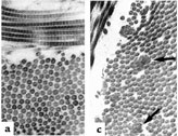
of JohnGohn (a) Normal collagen fibrils are of uniform size and
spacing. (c) Fibrils from a patient with classical EDS may show
composite fibrils (arrows))
This inherited disorder is caused by gene mutations that decreases the
function of collagen within the body. Collagen is a protein that
provides structural support to connective tissues including skin,
tendons, ligaments, blood vessels, and organs (A.D.A.M Medical
Encyclopedia, 2010). A full life span is expected for individuals
with most types of EDS, but the daily functioning of some
individuals is greatly altered. Unfortunately, EDS is not a
well-known disorder among the physical therapy community. More often
than not, EDS will be a secondary diagnosis that will influence our
functional outcomes while treating an acute injury, pain or
instability. It is important for physical therapists to become more
aware of this disorder and modify treatments accordingly to allow
patients to reach their full rehab potential.
There are six recognized types of connective tissue disorders that
can manifest in individuals with EDS. The types are categorized by
specific gene mutations and the signs and symptoms of the connective
tissue disorders. The six types are: arthrochalasia, classic,
dermatosparaxis, hypermobility, kyphoscoliosis, and vascular. Three
of the most common types are described in Table 1 along with
physical therapy implications. Although variable by type, the most
common symptoms of any kind of EDS is joint hypermobility and
increased skin elasticity (Mayo Clinic Staff, 2010).

In many cases, EDS can be diagnosed through symptoms and family
history. Genetic testing, skin biopsies to analyze collagen, and
echocardiograms to check heart health and valves may also be
indicated at times to determine the type of EDS present (Mayo Clinic
Staff, 2010). Due to the decreased knowledge of EDS treatment,
physical therapists are in a position to be an important advocate
for a patient or client with EDS in both the outpatient and
inpatient settings. In the inpatient setting, it may be appropriate
to educate caregivers and/or nursing staff on proper bed mobility
and transferring of the patient as to avoid shearing and skin
tearing of the already weakened integumentary system. In the
outpatient setting, being aware of increased joint laxity is
important when attempting to regain limited ROM. Excessive end-range
motions should be avoided to prevent dislocations and joint pain.
Without the collagen protection to keep the joint capsule strong,
individuals with EDS may also develop early onset of arthritis and
joint pain (Mayo Clinic Staff, 2010). Grade I-II joint mobilizations
may be indicated for pain relief, but high grades should be avoided
to limit unnecessary stress on the joints. A focus on joint
stabilization exercises may help other surrounding muscles support
the unstable joint. Physical therapy can also help in the treatment
of arthritis through pain management and strengthening exercises to
help protect arthritic joints (Mayo Clinic Staff, 2010). In extreme
cases, braces may be used to stabilize joints to increase function
with daily activities.
Individuals with vascular EDS have a bigger risk of developing
complications secondary to the frailty of blood vessels and organs.
These complications can be life threatening and can include aortic
dissection, organ rupture, and uterine rupture. Pregnancy can be
dangerous for an individual with vascular type EDS due to the
increased stress on blood vessels and organs, thus increasing the
risk of the above complications (U.S. National Library of Medicine,
2011). Exercise modifications may be indicated when treating a
patient with vascular type EDS as to not over-stress blood vessels.
Vitals should be monitored during treatment and proper rest breaks
should be given based of the cardiac demands of the exercise. The
median life expectancy age is 48 years old among people with
vascular type EDS due to the severity of complications (Mayo Clinic
Staff, 2010).
People with Ehlers-Danlos syndrome require a variety of health care
professionals to be knowledgeable about specific signs, symptoms,
and complications of the disease to be able to provide a
comprehensive continuum of care. Physical therapists may sometimes
be the first health care professionals to really spend time with
patients to recognize symptoms of hypermobility and increased skin
elasticity. This allows us as physical therapists to advocate for
patients/clients with suspected EDS and properly refer to other
health care professionals.
For more information please visit the Ehlers-Danlos National
Foundation at www.ednf.org.
Last revised: November 20, 2011
by Sarah Meuler, DPT
References:
1) A.D.A.M Medical Encyclopedia.
(2010, November 7). Ehlers-Danlos syndrome. Retrieved November 13, 2011,
from PubMed Health: http://www.ncbi.nlm.nih.gov/pubmedhealth/PMH0002439/
2) Mayo Clinic Staff. (2010, April 20). Ehlers-Danlos syndrome. Retrieved
November 13, 2011, from MayoClinic.com: http://www.mayoclinic.com/health/ehlers-danlos-syndrome/DS00706
3) U.S. National Library of Medicine. (2011, November 7). Ehlers-Danlos
syndrome. Retrieved November 13, 2011, from Genetics Home Reference: http://ghr.nlm.nih.gov/condition/ehlers-danlos-syndrome


