|

Many drugs are poorly absorbed
through skin by passive diffusion alone. The use of topical agents
often requires vehicle formation or chemical penetration enhancers
that are potential irritants or sensitizers (1). Iontophoresis and
phonophoresis are methods of driving topically applied substances
across tissues by utilization of electric current or ultrasound,
respectively. These physical modalities offer methods for enhancing
the percutaneous absorption of selected drugs (2).

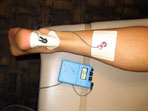
Iontophoresis is the use of electromotive force to enhance
percutaneous absorption of a drug or chemical. Iontophoresis usually
employs a direct current between 0.5 and 5.0mA2. The use of
iontophoresis has fluctuated over the years, partly due to concerns
about chemical burns of the skin that can accompany iontophoresis
treatment and the need for additional research demonstrating the
efficacy of the technique (3).
Many studies have been conducted on the topic of iontophoresis;
however, few have succeeded in accurately quantifying the useful
data and than applying the statistics to the clinical setting. For
example, Costello et al. (3) provided general information pertaining
to iontophoresis including: advantages/disadvantages, influencing
factors, applications and history. It was a thorough review of
previous research which demonstrated the need for further research.
However, his research would have better validity if he used
statistical analysis to support his conclusions. Therefore, the more
quantitative analysis rather than the qualitative analysis would
provide more meaningful information. Costello et al. (3) concluded
that the use of iontophoresis in medicine is likely to increase, but
questioned the extent to which physical therapists will be involved
in this expansion of usage. This secondary reference provides a good
foundation for researching the subject of iontophoresis as well as
produces results that are supported and generalizable.
The technical characteristics and mechanisms of action of
iontophoresis can be used as sources of research for the future. Why
does iontophoresis work the way it does and what would happen if any
conditions were changed? Kassan et al (2) reviewed the underlying
principles, current status, and potential of iontophoresis. Previous
research is adequately reviewed; however the lack of standardized
experimental conditions with respect to intensity of current,
frequency, waveform, on/off ratio of current, tissue pH and failure
to use adequate controls made it difficult to assess many of the
previous studies. The lack of gold standards in the use of
iontophoresis and the numerous varying methods and measurement
techniques made it difficult to compare/contrast research findings.
Lack of control has a large effect on the conclusion of findings
(2). Selection of homogenous groups was rarely performed in any
studies reviewed by Kassan. The subjects had different types of
injuries, damages to varying tissues, and reported various levels of
pain prior to onset of study. The level of control increased in
terms of treatment application. Each study had clear specifications
for how to the treatment was to be administered (4-6). Of course,
each research design differs slightly, but parallels can be drawn
and general conclusions can be made despite such differences.
Various levels of measurement can also make it difficult to
compare/contrast results from different studies. Ordinal data is
most often collected by means of some type of self perceived pain
scale (3-6). This indirect measurement scale always produces some
level of threat to validity, since each individual perceives pain
differently and levels of pain tolerance vary from person to person.
Therefore, the researcher must take these threats into consideration
when critiquing his/her methods, findings, and conclusions.

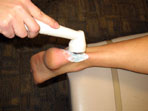
Phonophoresis refers to a specific type of ultrasound application in
which pharmacological agents such as corticosteroids, local
anesthetics and salicylates are introduced (7). Ultrasound is
primarily used for its ability to deliver heat to deep
musculoskeletal tissues such as tendon, muscle, and joint
structures. The depth of penetration is roughly inversed related to
the frequency (8). The therapeutic effects of heat likely involve
increased regional blood flow, increased soft tissue extensibility,
and decrease pain and muscle spasm (8). The mechanical properties of
ultrasound are less well defined but are believed to alter cellular
permeability and metabolism (9). The tissue response to these
non-thermal effects may be important in the promotion of wound
healing (9).
Due to their close relationship in methodology, phonophoresis and
ultrasound are often compared in terms of effectiveness. Klaiman et
al (7) looked at the differences in pain response after the
previously mentioned treatments. Therefore, a threat to internal
validity was present in spontaneous healing. The effects of no
treatment were not taken into comparison; therefore, previous
literature was relied upon to provide evidence in support of
treatment over no treatment.
Klaiman et al. (7) conducted a thorough literature review and
pointed out that the efficacy of phonophoresis has not been
conclusively established. In looking at both ratio and ordinal data,
these studies have a number of methodological shortcomings,
including inadequate blinding, lack of randomization, and
questionable assessment of pain relief (7). Nonhomogeneity of
subjects was a concern for control, but all of the testing
procedures were adequately controlled. No differences were found
between the phonophoresis group and the ultrasound group in
measurements of pain (visual analog system) or tolerance to
application of a pressure algometer.
From the review, one can make the case for the need for controlled
clinical studies comparing the efficacy of iontophoresis and
phonophoresis. This need is clearly shown by the lack of total
research conducted on the topic as well as the lack of measured
physiological changes associated with each modality. Research
questions that need to be answered relate to the actual depth of
drug penetration, the appropriate concentration of drug, the type of
vehicle preparation (cream, gel or ointment), the ultrasound
frequency, and the ultrasound mode (continuous or pulsed) (7).
In the research available that does compare/contrast iontophoresis
and phonophoresis, the lack of quantification seems to be evident.
Most measurements are done in relation to pain reduction, rather
than depths of muscle/tissue penetration. Conclusions that suggest
that iontophoresis and phonophoresis are promising methods of
enhancing topical delivery of both dermatological and
non-dermatological drugs (2) are clinically applicable and important
but further research should be conducted to search for more
statistical, physiological evidence. It has indeed been proven
through numerous research findings that both iontophoresis and
phonophoresis are effective methods of treating musculoskeletal
disorders. However, a future study should be designed to quantify
the actual differences of these treatments by using them in ways
that simulate realistic clinical situations.
Last revised: March 8, 2009
by Jennifer Hill, MPT, CSCS
References
1) Ronade VV. Drug delivery systems: transdermal drug delivery.
J Clin Pharmacol. 1991;31:401-18.
2) Kassan DG, Lynch AM, et al. Physical enhancement of
dermatological drug delivery: Iontophoresis and phonophoresis. J AM
Acad Derm. 1996;34:657-66.
3) Costello CT, Jeske AH. Iontophoresis: Applications in transdermal
medication delivery. Phys Ther. 1995;75:554-563.
4) Maloney M, Bezzant JL, et al. Iontophoresis administration of
lidocaine anesthesia in office practice. J Dermatol Surg Oncol.
1992;18:937-40.
5) Russo J, Lipman A, et al. Lidocaine anesthesia: comparison of
iontophoresis, injection, and swabbing. Am J Hosp Pharm.
1980;37:843-7.
6) Kennard CD, Whitaker DC. Iontophoresis of lidocaine for
anesthesia during pulsed dye laser treatment of port-wine stains. J
Dermatol Surg Oncol. 1992;18:287-94.
7) Klaiman MD, Shrader JA, et al. Phonophoresis versus ultrasound in
the treatment of musculoskeletal conditions. Med Sci Sports Exerc.
1998;30:1349-1355.
8) Lehman JF, DeLateur BJ, et al. Heating of joint structures by
ultrasound. Arch Phys Med Rehabil. 1968;49:28-30.
9) Dyson M, Pond JB. The effect of pulsed ultrasound on tissue
regeneration. Physiotherapy. 1970;56:136.
|


